Modeling is important in every field, and the better the model, the better the modeling. A model train without wheels isn’t going to take you very far. Little can be learned from a subpar model that lacks the complexity of the real thing—especially a model of something as complex as the human brain.
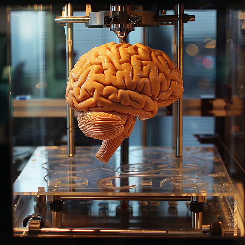
Thanks to a new method of 3D-printing, brain modeling has taken a major step forward, and as a result, whatever was once possible in neuroscience—and that encompasses a lot—may soon be put to shame. Some remarkable new research at the University of Wisconsin Madison, as discussed in the journal Cell Stem Cell, shows, for the first time, scientists 3D-printing functional brain tissue (1).
This research came out of UWM’s Waisman Center, which is dedicated to “Advancing knowledge of human development, developmental disabilities, and neurodegenerative diseases.”
This breakthrough in 3D-printing will likely lead to new advances in all of the above and beyond, including the potential development of new drugs for the full range of brain diseases. Such substantial potential for new research is made possible by a revolutionary aspect of this study: the communicative interactivity of the printed cells (1).

Functional Brain Cells Are Chatty
What makes brain cells functional? And why is isolated brain tissue not that interesting or useful in a model or the real thing?
Think of brain cells like people at a party. If no one is talking, and everyone is shuffling around on their own, eating appetizers awkwardly, that’s not a fun or functional party. In fact, it’s a giant bummer. The same goes for brain cells; they need to interact. A brain in which the cells don’t interact is no brain at all.
The tissues created at the Waisman Center are big news because they’re capable of forming neural circuits and functional connections—the kinds of hookups that make a brain a cohesive organ and not a lump of uncommunicative cells. That’s the good kind of party, where everyone is talking and joking and laughing, and it’s the kind of brain tissue that mimics the wonders of a real brain (1).
3D Bioprinting: A Small But Significant Change
Sometimes, even in brain science, one small change is all that is needed for a significant breakthrough. Here, that small change involved switching up the typical method of 3D bioprinting, which usually involves printing and stacking in vertical layers. As described in Cell Stem Cell, the new method involves
“printing one layer or band next to another horizontally rather than the traditional way of stacking the layers vertically. These specially designed 3D neural tissues can be maintained by the conventional culture systems and are amenable to easy live-cell imaging and electrophysiological recording, providing a new platform for examining human neural networks under physiological and pathological conditions.”
By switching to horizontal, everything changed. The vertical layers were stacked too tightly, preventing growth, connection, and functionality. But thanks to the new horizontal method, the neurons had room to “breathe.” This method allowed neurons the space to grow into each other and interact—just like “real” brain tissue (1).
Such interactions make a brain work properly, doing everything from remembering what you did last summer to deciding what to order for lunch. The interconnectivity of brain tissue also allows a brain to adapt after an injury.
Since these 3D-printed cells have the full, conversational functionality and versatility of brain cells, they should prove essential in research into a variety of brain diseases and neurological conditions. Disease aside, the artificial but authentic neurotissue will also offer insight into how healthy brains operate by forming networks of neurons (1).
A Greatly Improved 3D Printing Model of the Brain
Since the 3D-printed cells work just like real brain cells, they are far superior to existing models of the brain. Such methods, which may soon be passe, include MRIs and brain organoids.
A tried and true method of imaging and modeling the brain is MRI (magnetic resonance imaging). When doctors need a closer look at someone’s brain, MRI gets the call, and such imaging has been invaluable in diagnosing brain damage and other neurological conditions.
But the MRI may be surpassed soon. Research has found that a process called two-photon polymerization (2PP) 3D printing can produce “phantoms”—sample tissue that mimics genuine brain tissue. This is just one example of how 3D printing will revolutionize neuroscience by creating better models. (2).
Another method that may go the way of the dodo is the brain organoid—a small, three-dimensional organ grown to mimic part of the brain. 3D-printed brain tissue has great advantages over organoids in that
“the printed tissue allows the incorporation of different neuronal subtypes such as cortical glutamatergic neurons, cortical GABAergic interneurons, or striatal medium spiny neurons at defined proportions, which is difficult to achieve via organoids or other printing methods to date.” (1)
In essence, the 3D-printing allows for the creation of specific and specialized tissue, which can be made to order.
3D Printing for Brain Research: Custom Brain Tissues for Neurological Studies
Indeed, one of the primary advantages of this new method of 3D printing is the ability to print specific types of brain tissue. This allows researchers maximum flexibility in their brain studies, allowing them to produce the type of brain tissue needed to study Down syndrome, Alzheimer’s, autism spectrum disorder, or Chronic Traumatic Encephalopathy (CTE). The brain tissue can be produced as needed to match the problem at hand with precision that, until now, just hasn’t been possible (1).
Different types of brain tissue, with full communicative potential, can be created like neural legos, with the same ability to build connections as our brains. These neural networks offer a smorgasbord of learning opportunities. Previous generations of neurologists could only dream of having such realistic models of actual living brains, but today’s neurologists will be able to make such dreams a reality (1).
The Future Starts Now
With the development of such authentic, functional brain tissue, our understanding of the brain is sure to increase, perhaps exponentially. This could mean fantastic advances in the study of brain diseases and brain injuries in the near future. Also, how the most mysterious organ of them all really works will become less and less of a mystery, as new models provide long sought-for answers.

Elevate Your Medical Communications with DKMD Consulting
Medical advancements are occurring at a breathtaking pace, and conveying these breakthroughs effectively to your audience is the lifeblood of your business. At DKMD Consulting, we specialize in transforming complex medical innovations, like the recent strides in 3D-printed brain tissue, into clear, impactful communications that resonate with clinicians and patients.
Understanding the intricacies of scientific research can be challenging. Our team excels at distilling this complex information into digestible, engaging content that informs and inspires action. Whether you want to educate your clientele about new treatment methodologies or showcase cutting-edge research, DKMD Consulting crafts the narrative that aligns with your goals.
Partner with us to enhance how your business communicates about health and science:
Expert Medical Writing: Benefit from our expertise in medical copywriting to ensure your communications are scientifically accurate and compelling.
Content Strategy Development: Let us help you build a content strategy that effectively communicates the value of the latest medical advancements to your audience.
Educational Materials and Resources: Engage your clients with beautifully crafted educational materials that simplify complex concepts.
Step forward with DKMD Consulting and ensure your communications reflect the sophistication and potential of modern medical science.
Contact us today to learn how we can help you articulate the future of healthcare.
References
- Yan Y, Li X, Gao Y, Mathivanan S, Kong L, Tao Y, Dong Y, Li X, Bhattacharyya A, Zhao X, Zhang SC. 3D bioprinting of human neural tissues with functional connectivity. Cell Stem Cell. 2024 Feb 1;31(2):260-274.e7. doi: 10.1016/j.stem.2023.12.009. PMID: 38306994; PMCID: PMC10883639.
- Woletz, M., Chalupa-Gantner, F., Hager, B., Ricke, A., Mohammadi, S., Binder, S., Baudis, S., Ovsianikov, A.,DKMD Consulting Windischberger, C., & Nagy, Z. (2024, January 07).
Toward Printing the Brain: A Microstructural Ground Truth Phantom for MRI